
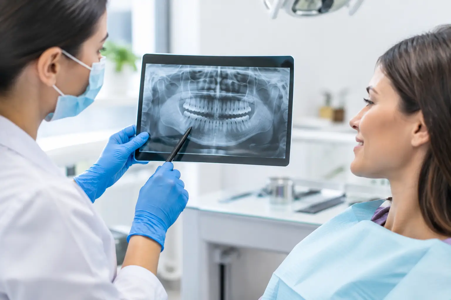
A tooth can look perfectly fine from the outside while a cavity develops between it and its neighbour, or an infection forms quietly beneath an old filling. That is why patients often ask, are dental x-rays safe? The reassuring answer is that, when they are clinically needed and taken with modern equipment, dental X-rays use a very small amount of radiation and are an important part of protecting your oral health.
At Smile District Dental, we understand that the word “radiation” can feel worrying, particularly if you are bringing in a child, are pregnant, or have had several medical scans before. Your dentist will recommend X-rays only when they can provide information that cannot be gathered properly through an examination alone. The goal is simple: find concerns early, plan treatment accurately and help you avoid bigger, more uncomfortable problems later.
Are dental X-rays safe with modern technology?
Modern dental imaging is designed to keep exposure as low as reasonably achievable while still producing a clear, useful image. Digital dental X-rays generally require far less radiation than older film systems. They also allow the dentist to view the image straight away, enlarge areas of concern and plan treatment with greater precision.
The radiation from a routine dental X-ray is low and adds only a small amount to the natural background radiation we all receive from everyday surroundings. The exact amount varies according to the type of image needed. A small X-ray of a few teeth uses less exposure than a panoramic image of the jaws, while a 3D cone beam scan is reserved for situations where detailed planning is genuinely required, such as implants, complex wisdom tooth removal or certain root canal treatments.
Low exposure does not mean no care is needed. Dental teams follow strict safety processes, use appropriately maintained equipment and select the smallest, most targeted image that answers the clinical question. If an X-ray will not change the way your dentist assesses or treats your teeth, there is no benefit in taking it.
Why an examination alone cannot show everything
Your dentist can see a great deal during a check-up: the condition of your gums, visible decay, cracks, wear, plaque buildup and changes to your soft tissues. But many dental problems begin where the eye cannot see.
X-rays can reveal decay between teeth, infection around tooth roots, bone loss linked with gum disease, impacted wisdom teeth, problems beneath crowns or fillings, and the position of developing adult teeth in children. They are also useful when a tooth becomes painful after an injury, even if it has not chipped or changed colour.
Early detection matters because treatment is often simpler when a problem is small. A tiny cavity may need a modest filling. Left undetected, it can progress towards the nerve of the tooth and lead to pain, root canal treatment, a crown or even extraction. The trade-off is considered carefully: a small, controlled amount of radiation may prevent a far more serious oral health issue from developing unnoticed.
How often should dental X-rays be taken?
There is no one schedule that suits every patient. The right timing depends on your age, oral health history, risk of decay, gum health, symptoms and the quality of any previous images available to your dentist.
Someone with healthy teeth, low decay risk and no symptoms may not need bitewing X-rays at every check-up. A person with frequent decay, dry mouth, gum disease, extensive restorations, new dental pain or a history of treatment between visits may benefit from imaging more often. Your dentist should be able to explain what they are checking for and why the image is recommended now.
Children may need X-rays at certain stages because their mouths change quickly. Images can help monitor adult teeth as they develop, identify cavities between baby teeth and assess whether there is enough room for teeth to erupt normally. This does not mean children are X-rayed routinely without reason. The same careful, individual approach applies, with exposure kept as low as possible.
What types of dental X-rays might be recommended?
The type of X-ray depends on the concern your dentist is investigating. Bitewing X-rays show the crowns of upper and lower back teeth together and are particularly helpful for spotting decay between teeth. Periapical X-rays show the whole tooth, including its root and surrounding bone, and may be used for pain, infection or root canal assessment.
A panoramic X-ray captures both jaws in one image. It can help assess wisdom teeth, jawbone health, missing teeth, tooth development and certain jaw concerns. For more complex planning, a cone beam CT scan provides a three-dimensional view. It gives valuable detail for procedures such as dental implants, but because it involves more radiation than a small intraoral X-ray, it is not used as a routine screening tool.
Dental X-rays during pregnancy
If you are pregnant or think you may be, tell the dental team before any imaging is taken. Urgent dental concerns should not be ignored during pregnancy, particularly infection or significant pain. In many cases, a dental X-ray can be taken safely when it is necessary for diagnosis and treatment planning, using modern digital technology and appropriate protective measures.
Elective imaging can sometimes wait if there is no urgency. Your dentist will weigh the reason for the X-ray, your stage of pregnancy and whether delaying it could allow a problem to worsen. Open communication helps your care stay both safe and comfortable.
What you can expect at your appointment
Taking a dental X-ray is usually quick. For small intraoral images, a sensor is placed gently inside your mouth while you bite down briefly. If gagging or anxiety is a concern, let the team know before you start. There are practical ways to make the process easier, including taking breaks, adjusting positioning and explaining each step before it happens.
For a panoramic image, you stand or sit still while the machine moves around your head. It does not touch your teeth and is over within seconds. Your dentist will then review the images with you in plain language, showing you what they can see and discussing whether any treatment or monitoring is needed.
Questions worth asking your dentist
Feeling informed can make imaging far less stressful. You are always welcome to ask why an X-ray is being recommended, what type of image is needed, whether a recent image can be used instead, and how the result may affect your treatment plan.
If cost is a concern, ask for an estimate before proceeding. If you have previous X-rays from another clinic, mention this as well. Existing diagnostic images may still be useful, depending on their age, quality and the issue being assessed.
When delaying an X-ray may create a bigger problem
It can be tempting to decline an X-ray because there is no pain, or because a previous dental visit felt uncomfortable. Yet dental disease is often quiet in its early stages. Decay between back teeth may not hurt until it is advanced. Bone loss from gum disease can progress gradually. An infection at the root of a tooth may only become obvious when swelling or severe pain develops.
A caring dental team will never pressure you into imaging without an explanation. However, when an X-ray is clinically indicated, it gives your dentist a clearer view of your health and allows you to make a genuinely informed choice about next steps. That clarity is especially valuable before major treatment such as crowns, implants, wisdom tooth extraction or full-mouth rehabilitation.
If you have been putting off a check-up because you are concerned about X-rays, start with a conversation. A gentle appointment, clear answers and an individual plan can turn an unknown into a manageable part of looking after your smile – and your confidence.